 Overbeck/L2phase transition in the C
21monolayer
Overbeck/L2phase transition in the C
21monolayer
SAXS is a small-angle scattering laboratory technique in which photons (wavelength between 0.1 and 0.2 nm) are elastically scattered from a sample due to differences in electron density between the solute and the solvent.
Small angle scattering of x-ray is observed from almost all kinds of matters, and it is widely used in structural studies of non-crystalline materials at relatively low resolution. The term "small angle" here refers to the angular range within a few degrees (range between ~ 0.1° and 10°), containing structural information on the order of approximately a nanometer to submicrometers.
The advantage over crystallography is that the samples need not be crystalline, must not be too thick and features on the nanometer length scale can be examined.
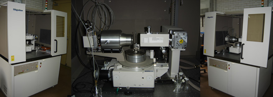
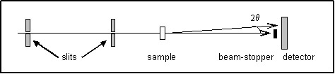
SAXS experiments are usually performed in transmission mode (Fig.1). The maximum scattering
intensity in this mode, from any arbitrary material, is obtained for a sample thickness more less 1/μ(λ), where
μ(λ) is the linear absorption coefficient.
Applications
This technique is employed to study nanostructured materials, developing of new materials, to derive size and shape parameters of large molecules which can not be determined by other methods (such as microscopy or X-ray diffraction), for studying the morphology of complex aggregates, the determination of the persistence length (a measure of flexibility) of polymers in solution, for the domain structure of solid polymers, for the determination of the specific inner surface of powders, long-range order of liquid crystals, analysis of heterogeneous materials, characterization of fractal structures, micro porous materials, etc. In structural biology this technique complements crystallographic structural analysis, which requires hard-to-get high quality crystals of macromolecules, and is one of a few structural techniques for studying proteins in solution such as those in unfolded states or simply those whose crystallization conditions have not been determined. Biological fibers such as skeletal muscle are quasi-crystalline but not as well ordered as crystals, thus giving relatively broad diffraction peaks mostly at small angles. Many biological lipids exist as vesicles or liquid crystals, physical states that are rather poorly ordered. Micellar structures and synthetic polymer materials are often studied with this technique.